


Principle for Ametropic Correction of iLASIK Surgery
Procedures of Surgery
Introduction to Characteristic of Various Equipments
Selection of Surgery (Indication and Price)
Dos and Don'ts before and after Surgery

High-level Intraocular Lens Measuring Instrument and Operation
1. Intraocular Lens Measuring Instrument (IOL Master) Zeiss IO
Zeiss IOL Master improves patients’ vision and quality of life after IOL implantation.IOL Master provides usable biological formulas for calculating IOL: SRK II,Hoffer Q,Holladay,SRK/T,Haigis
Advantages:
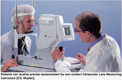
High precision: Measure patient’s axial length of eyeball along Visual Axis, from surface of lacrimal film to RPE (retinal pigment epithelium) layer.
Non-contact technology: Would nor damage cornea for local anesthesia, no need to dilate pupil and press cornea, and avoid the possibilities of cross infection and damage to cornea.
Keep patient’s safety and provide comfortable measurement for patients: No need to anesthetize, simple and rapid operation, would not feel uncomfortable for pressing cornea;
The results of measurement would not be different with operators: The clinical trials proved that results of measurement would not be changed with operators.
Time saving:The patients don’t have to move to another instruments for desired data, the three-in-one design (K Reading, Axial Length & Anterior Chamber Depth and WTW measure) has greatly shortened the time taken to measure and provides immediate IOL calculation after obtained the desired data. Compared with ultrasonic technology, that three-in-one design has greatly reduced the time used in surgery by 25%.
Can be used for several patients simultaneously, and build independent data bases.
Compared with 4.0 version, the current 5.0 upgrade version can enhance the effective detection rate centered on relatively mature cataract.For patients who have gone through LASIK surgery, the data can be precisely measured and calculated by Haigis program to increase the precision without K value before surgery.
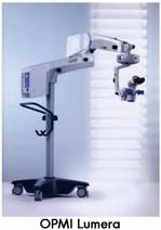
2. Operation Microscope (OPMI Lumera)
The optical system with technological structure of Zeiss microscope innovates from old ones and has multi functions, which adopts the new depth of field design of Deep View. This kind of microscope can provides optimal transmission between magnification system and brightest light, increase the resolution and contrast. With built-in three-dimensional assisting microscope, it can provide identical working conditions and independently adjust focus and magnification. Moreover, the assisting microscope of it would not weaken the light of main surgeon. No special setting is needed if moving from left eye to right eye, only simply rotates the assisting microscope and goes on the surgery, which showed that it is the optimal operation microscope. Besides, it has xenon lamp illumination, high contrast and true color image presentation.Built-in slit source, obtain high contrast through lens reflection
Easily adjustable handle design based on ergonomics;
Totally achromatic optics;
BrightFlex light source system, bright, strong sensitivity for iris;
Many optional parts
